In medical imaging, clarity and precision are not just technical goals—they’re essential for accurate diagnosis and effective patient care. Even the most advanced X-ray machines can’t compensate for improper positioning. That’s where x ray positioning aids become indispensable. These tools help radiologic technologists ensure the patient and the targeted anatomy are aligned correctly, reducing image distortion, minimizing retakes, and enhancing diagnostic reliability.
Understanding the Role of X Ray Positioning Aids
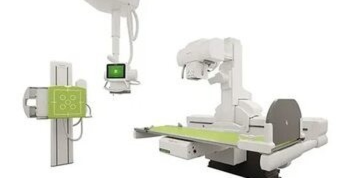
What Are X Ray Positioning Aids?
X ray positioning aids are specialized tools and supports designed to hold a patient or a body part in a specific position during X-ray procedures. They vary widely depending on the imaging need—whether it's for the spine, limbs, head, or torso. These aids help maintain stillness, minimize discomfort, and support anatomical accuracy.
Common types of positioning aids include:
Foam blocks and wedges
Sandbags and straps
Lead markers and grids
Radiolucent sponges
Immobilization devices
Each type is tailored to a specific imaging requirement and helps create consistency in imaging across patient populations.
How They Work in Sync with Radiologic Protocols
Radiology departments follow strict imaging protocols to ensure reproducibility and minimize radiation exposure. Positioning aids are an integral part of these protocols. They:
Stabilize anatomy to prevent motion blur
Align the subject with X-ray beam angles
Support symmetry in bilateral imaging
Help adhere to standard views (e.g., AP, lateral)
The Direct Impact of X Ray Positioning Aids on Image Accuracy
Minimizing Motion Artifacts
Motion is the biggest enemy of image sharpness in radiology. Even slight movements can result in blurred images that compromise diagnostic clarity. By holding the patient still, x ray positioning aids dramatically reduce motion artifacts, especially in pediatric and geriatric imaging, where patient cooperation may be limited.
Achieving Consistent Anatomical Views
Positioning aids are vital for creating reproducible and standardized views. Whether it’s a chest X-ray or a lateral view of the cervical spine, these aids help maintain the correct angles and distances between the anatomy and detector, enhancing consistency and accuracy in imaging.
Reducing Repeat Exposures
Improper positioning often leads to repeat X-rays, which not only delay diagnosis but also increase radiation exposure. Using high-quality positioning aids helps get it right the first time, improving workflow and safeguarding patient safety.
Types of X Ray Positioning Aids That Deliver the Best Results
Foam Positioning Aids
Foam blocks and wedges are radiolucent and ideal for positioning limbs, heads, and trunks. Their non-slip surface and contoured design ensure stability without interfering with image quality.
Radiolucent Sponges
These are lightweight and do not show up on X-rays, making them ideal for accurate positioning without any image obstruction. Their varied shapes accommodate different anatomical needs.
Sandbags and Straps
Useful for immobilizing arms, shoulders, or legs, sandbags and straps provide weighted resistance, especially when patients have difficulty holding positions.
Pediatric Immobilizers
Children present unique imaging challenges due to movement and discomfort. Pediatric immobilizers with soft padding and adjustable sections allow for comfortable, secure positioning while ensuring clear imaging.
Positioning Markers and Indicators
Radiographic markers indicate patient orientation (e.g., L for left, R for right) and play a vital role in diagnostic interpretation. Combined with positioning aids, they offer complete context in each image.
Benefits of Using X Ray Positioning Aids in Clinical Practice
Improved Diagnostic Confidence
Clearer images lead to better diagnostic interpretation. Radiologists can detect abnormalities like fractures, tumors, or infections with more confidence when the images are well-positioned and artifact-free.
Enhanced Patient Experience
Patients benefit from shorter procedure times, reduced discomfort, and fewer repeat exposures when positioning is correct the first time. This improves overall satisfaction and compliance, especially in repeated imaging sessions.
Higher Departmental Efficiency
Fewer image retakes mean smoother workflows, reduced costs, and optimized use of radiologic equipment and staff. This is particularly valuable in high-volume settings like emergency rooms and outpatient centers.
Real-World Applications: Where Positioning Aids Make a Difference
Orthopedic Imaging
In musculoskeletal exams, precision is essential. Positioning aids ensure bones and joints are correctly aligned for clear imaging of fractures, joint spaces, and soft tissue injuries.
Chest and Thoracic Imaging
Proper alignment of the thorax prevents oblique shadows or rotated views that can obscure pathology. Chest boards and foam wedges help achieve the perfect PA or lateral chest view.
Dental and Cranial Radiography
Intraoral and panoramic imaging require stable jaw and head positioning. Bite blocks and headrests improve precision while reducing patient fatigue and movement.
Operating Room Imaging
Portable imaging during surgeries demands quick yet accurate positioning. Radiolucent aids allow for intraoperative imaging without moving the patient extensively.
How to Choose the Right X Ray Positioning Aids
Consider the Patient Population
Pediatric, bariatric, or geriatric patients require specialized aids. Pediatric aids need to be soft and adjustable, while bariatric tools should be wider and more durable.
Match the Aid to the Exam Type
A headrest won’t work for a pelvic X-ray. Know which aid best supports the region being imaged. For spinal alignment, foam wedges and spine boards work best.
Focus on Infection Control
Choose aids with wipeable, antimicrobial surfaces. In high-traffic departments, this is essential to prevent cross-contamination.
Look for Radiolucent Materials
A good aid should not show up in the image or distort anatomical views. Radiolucent foams and plastics are the industry standard.
Maintenance and Best Practices
Regularly inspect for wear and tear
Disinfect after every use
Replace compressed or damaged foam aids
Train staff in correct usage to ensure consistency
Final Thoughts: Elevating Image Quality with the Right Tools
Using x ray positioning aids is not just about holding a patient still—it’s about ensuring diagnostic excellence, patient safety, and operational efficiency. As imaging technology advances, the demand for precise, repeatable, and clear diagnostic results grows. Radiology departments that invest in high-quality positioning aids are setting a standard for accuracy and care that directly benefits both clinicians and patients. Embracing these tools means every X-ray taken is one step closer to clinical clarity and diagnostic confidence.